Monday, November 16, 2009

Observation 5

(11/12/09)
OBSERVATION 5

The first part of naked eye observation is visible in the glass tank is annelida (white spiral thread-like) at the surface of the glass tank and Cyclops (motile gray spots) in different area around Plant A & B.

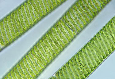
Under microscope of high power, I saw many multicellular Cyclops, even young stages of Cyclops swimming around Plant B. Here is MP4 of Lecane Rotifer, identified by Dr. McFarland, is compressed 1.35 megabytes with a playing time of 6 seconds. It is eating or swimming around zygnema. Zygnema's the bunch of green string of filaments.

MP4: Lecane Rotifer

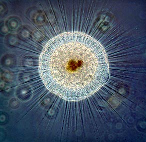
Many of Aspidisca (Paterson 119) are found at surface of the glass tank. It is more round and has a strange mouth as the beak. It has many flagella to move circular in the tank. See the MP4 video of Aspidisca (please excuse the wrong identity of discomporhia) below. It is compressed in 4.75 megabytes and it is under lightfield illumination at a magnification of high power with a playing time of 20 seconds.

MP4: Aspidisca

Noticed the picture of Coleochaete irregularis (Forest 89) from third week of observation is a family to this picture below of figure 1. It is Coleochaete orbicularis (Prescott 105) because of its filaments attached to the discs and it is branching out to stay.

Fig. 1

More picture of figure 2 below seems to look like one of Coleochaete because it has similar structure and it also has filaments attached to the dics except the cell structure is more rod-like. Other one on the picture of figure 3’s seems like first stage of Coleochaete orbicularis or it has lost some of filaments. (1624, 1625)

Fig. 2
Fig. 3


REFERENCES

Forest, Herman Silva. (1959). Handbook of Algae. The University of Tennessee Press. 89

Paterson, D.J. (2003). Free-Living Freshwater Protozoa. Manson Publishing: Washington D.C. 119 (fig 246).

Prescott, G.W. (1964). The Fresh-Water Algae. W.M.C. Brown Company Publishers. 105

Tuesday, November 10, 2009

Observation 4

(11/05/09)

INFORMATIONS
This week we are going to observe and find a general existing microanimal and microorganisms in the glass tank. We want know the numbers of the population in the glass tank, their growth process and densities. We are going to gather all observations before to write a final laboratory report on MicroAquarium.

OBSERVATION 4
My observations of this week on November 5th are that a few fresh-water communities are still growing and the population of rotifers, diatoms, desmids, nematodes and euglena are increasing. Though the population of diatom is dying and they are everywhere. See MP4: Annelida below. You will see the millions of thread-like algae and some dead microorganisms sprinkled around Plant A. The algae are growing plenty of “hair-like.” The Plant A & B are turning green to brown.

This microanimal is new to me. I saw several of annelida swarming around on Plant A & B. I took a MP4 video of it to see how it is moved by its several flagella. It is beneath the medium & high power of microscope. It is compressed in 10.3 megabytes and it is under lightfield illumination at a magnification of medium & high power with a playing time of 44 seconds. It is to be mobile and it moves slowly around like worm-like but in one place. It is completely covered by See that tail at the end of the video clip is a mouth. It is called Stylaria lacustris (Pennak 303), identified with help by Dr. McFarland and species found in the book, Fresh-Water Invertebrates of the United States by Robert W. Pennak.


MP4: Annelida


Spotted a new buddy in the glass tank I don’t think I have seen it before. It is an axial stellate, non-motile filamentous alga and it has two green (chloroplast) star-gazed in each cell. Lot of them like to float around Plant A and B. It is called Zygnema (Prescott 101). You can see the figure 1 of Zygnema at below and see more in Wim Van Egmond’s site: http://tolweb.org/tree?group=Annelida&contgroup=Bilateria.


Fig. 1: Zygnema

Others I don’t recognized its species, but it is fascinated to see that seems to be act like a gel on Plant A. See figure 2 below. I think it is Eudorina elegans (Egmond, 1994) because of its similar composition of cells and it looks like Volvox.

Fig. 2

It remains stationary and it seems it grows outward from the center. It is because of crowding among the filaments that forces the cells into abnormal circular shape. With help from Dr. McFarland and The book of Handbook of Algae shows it is Coleochaete irrregularis. (Forest 89). See Figure 3 below.

Fig. 3

The couple of bell stalk-like Vorticella stands out boldly on dying Plant A. (Patterson 113). See figure 4.

Fig. 4: Vorticella


The strange microanimal we could not identify in first week of observation reveals that it was just a young stage of Cyclop. You can see the flagella on its tail. It is starting to grow.

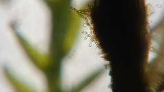
Fig. 5: The mystery finally revealed

OVERALL
Diatoms are dying due to its life period and it appears to be little crowding in the glass tanks. I supposed that is why they could not move around much to get nutrients. Since the results of 4 weeks of observation, more rotifers are continuing to grow. Counting the microorganisms, Cyclops and rotifers exceed more than other organisms in fresh-water pond from runoff from cemetery. The least microorganism is seed shrimp. I don’t see any more much of that.


REFERENCES

Egmond, Wim van. (1994). The Algae Archive of the Micropolitan Museum. Data Retrieved on 6 Nov. 2009.

Forest, Herman Silva. (1959). Handbook of Algae. The University of Tennessee Press. 89

Patterson, D.J. (2003). Free-Living Freshwater Protozoa. Manson Publishing: Washington D.C. 113.

Pennak, Robert W. (1989). Fresh-Water Invertebrates of the United States. John Wiley & Sons, Inc: New York. 303

Prescott, G.W. (1964). The Fresh-Water Algae. W.M.C. Brown Company Publishers. 101

Thursday, October 29, 2009

Observation 3

(10/29/09)

INFORMATIONS
This week we are expected to identify the fresh-water algae in the glass tank. A little lesson about algae, it likes to grow whatever there is nitrate and phosphate in the water. Only way to stop algae from growing by reducing these nutrients like “Beta” Food Pellet. That will stop an algae outbreak. Imagine trying to control an algae outbreak in fish tank… The term algae refer to microscopically small, unicellular organisms, that some form colonies and therefore reach sizes visible to the naked eye as tiny green particles (McFarland 107). The algae are known as photosynthetic aquatic Protista (McFarland 106). All of them are eukaryotic and they are pretty difficult to identify because it is so many of them are different and odd.

OBSERVATION 3
The third week of observation on Thursday, October 29th, I saw some of my old buddies are really growing up. I saw an adult Cyclop with an egg sac. Apparently the other egg sac had been hatched somewhere. I also saw young Cyclops swimming around. There are more blue-green cyanobacteria everywhere near the muck. Nematodes (flatworms), were identified by Dr. McFarland, are growing bigger and longer than before. They tend to swarm around Plant A & B.

I took a video of seed shrimp from last week on Observation 2, see what it is actually looks like beneath the high power of microscope. See MP4: Seed Shrimp below. It is compressed in 1.73 megabytes and it is under lightfield illumination at a magnification of high power with a playing time of 23 seconds. It appears to be immobile or mobile but in one place. It has shells and some more of flagella around its legs.

MP4: Seed Shrimp


Other adult fresh-water, multicellular organism I first saw is a magnificent rotifer on the muck and it was showing off its long finger cilia. First spooky as it looks but it contains 3 layers of digestion, muscle, and invertebrate shells. It has a full digestive system and it uses the long finger cilia to move food into its mouth and manipulate the water around them in order to move. The fresh-water microorganism is known as Limnias, Setphanoceros Eichornii (Plaskitt 219). See Mp4: Limnias at below. Compressed in 8.43 megabytes. Time: 1 minute, 29 seconds.

MP4: LIMNIAS


There are more rotifers on Plant A & B. There are 4 types of rotifers found in my glass tank. Other rotifer I saw in the tank is known as Philodina (Plaskitt 194). There are trap planet, diatoms - Bacillariophyta (Rainis et cal 116), Euglena helicoideus (Rainis et cal 283), desmids - Conochiloidses & Conochilus (D. Smith 179), Spondylosium (G. Smith 333) in the tank.

See Philodina in the MP4 file (Quickplayer 7) at below and it is compressed in 13 megabytes. It is at a magnification of medium and high power with a playing time of 1 minute, 41 seconds. Philodina: You can see the movement of short cilia on the margins of its mouth when it comes out and spread its mouth (G. Smith 192).

MP4: Philodina


Other good-ole organisms are doing their job to take care of my glass tank: see figure of diatoms and spondylosium at the bottom. Diatoms, to specifically identify it are difficult, but I think it as Mastogloia from the book, The Fresh-Water Algae (Prescott 243). It moves to left and right in slow motion or none at all. It is autotrophic in nature.


Fig. Diatoms & Spondylosium


REFERENCES

McFarland. (2009) General Botany 111 2009. Retrieved data by 27, Oct. 2009. http://botany1112009.blogspot.com/

Plaskitt, F.J.W. (1926). Microscopic Fresh-Water Life. Chapman & Hall, LTD: London. http://www.archive.org/details/microscopicfresh029632mbp. 194, 219

Prescott, G.W. (1964). The Fresh-Water Algae. W.M.C. Brown Company Publishers. 243

Rainis, Kenneth G. & Russell, Bruce J. (1996). Guide to Microlife. Franklin Watts: Danbury, Connecticut. 283

Smith, Douglas Grant. (2001). Pennak’s Freshwater invertebrates of the United States 4th ed. John Wiley & Sons, IAC: New York. 179

Smith, Gilbert M. (1950). The Fresh Water Algae of the United States 2nd ed. McGraw Hill book Company: New York. 333

Tuesday, October 27, 2009

Observation 2


INFORMATIONS
The plant A and the plant B that are in my glass tank which I was not sure at first, but more information comes from this link: http://botany1112009.blogspot.com/. The plant A is Amblystegium varium (Hedw). It comes from moss and it is collected in partial shade from the Natural Spring at Carters Mill Park on Cater Mill road.

The plant B is Utricularia vulgaris L. Flowering. It comes from the south shore of Spain Lake on Camp Bella Air road from east Sparta of Tennessee. It was grown in water tanks outside of greenhouse at Hesler Biology Building at the University of Tennessee.

Once a week I will have to add one small food for organisms in my glass tank. It is called "Beta" Food Pellet. It is made by Ocean Nutrition - Aqua Pet Americas from 3528 W. 500 S. in Salt Lake City of Utah. Its ingredients are fish meal, wheat flower, soy meal, krill meal, minerals, and vitamins.


OBSERVATION TWO
The second observation of my microaquarium on Thursday, Oct 22nd, I saw a few of the same organisms from last week. I saw Cyclop (Eucopepoda) with big black or dull-grayish egg sacs. Its one black eye located in the middle of the head. It swims jerkily as happily around the Plant A. The egg sacs show that it is a female organism and it is "free-living copepods." It looks like a grayish, elongated body and it have many flagellum to swim around. It prefers to be in quiet waters. There are several more of them in the glass tanks. Take a look at two images at bottom of Cyclop with egg sacs and a young stage of Cyclop.


Other new organism I didn't see in first observation, it looks like it comes from a family of Cyclop, but if you take a closer look at 3 images at bottom. It has a usual shape of blotted black eye unlike Cyclop's and it has arms like lobster. The problem is Cyclop have a long tail of the body, this one doesn't have any. They like to hang around the plant B.


I observed some of organism like the green alga, spirogyra, and an up-closed image of blue-green cyanobacteria at below. Some of new organisms I saw and identified by "Guide to Microlife" by Rainis and Russel: threadworms, flatworms (Stenostomum leucops), rhyziod, rotifera & seed shrimp (ostracods). See images below from top to bottom in order: threadworms, flatworms, cyanobacteria & seed shrimp.


This peculiar image of green alga (plant A) at the bottom shows some color of red and invisible linear roots. It is called filamentous rhizoid (rhyziod). Rhizoids are multicellular and it serves only to anchor to plants.


I can see its helix-shaped by cocci lined up spirally. That is spirogyra. It is filamentous, non-motile, and they lack flagellated cells. Its color pigment shows that it contains chloroplasts.

There is lot of going on in my microaquarium than before. Most of organism has grown bigger and more noticably in concise shape. Compare in first observation in Oct. 14th I used 4x and 10x to observed organims in the tank; I saw only one Cyclop, lots of zigzags cells, filamentous string-like spirogyra & cyanobacteria.


REFERENCES

Cook & McFarland. (1995). General Botany 111 Lab. Manual. Unit 16.

McFarland. (2009) General Botany 111 2009. Retrieved data by 20, Oct. 2009. http://botany1112009.blogspot.com/

Rainis, Kenneth G. & Russell, Bruce J. (1996) Guide to Microlife. Franklin Watts: Danbury, Connecticut. p. 179, 204, 209, & 230

Raven, Evert, & Eichhorn. (2005). Biology of Plant 7th ed. New York, New York: W. H. Freeman & Company Publishers. p. 348

Ward B., Henry & Whipple, George. (1918). Fresh-water Biology. New York: John Wiley & Sons, Inc. p. 335.

Friday, October 16, 2009

MicroAquarium Observation

(10/14/09)
I choose to do #10 on Lynnhurst Cemetery. I am curious what kind of microbes look like in the water and what it does. The water source is from Lynnhurst Cemetery's, located at Adair Drive, Knoxville, Tn. The water pool below spring is contaminated from run-off water from the cemetery. The muck from the water pool, 2/3 full surface water, and two other kind of mosses in the pocket of glass tank to be observed through the objective lens in low and medium power. There are various immobile and mobile organisms were identified during the lab: Cyanobacteria, Chlamydomonas, Euglena, Cyclops, and Spirogyra. There are so many of them I have not able to identified yet. Some organisms move in circular movement, some zigzag, many different of sizes, some remain stationary, others move really quick, and others move fro and back slowly. I created an image what I saw and some of organisms are strange to me.

Image created by Jo:

Desmids like Xanthidium? It looks like a star or spiky shape, bright as yellow/light green, stationary. (top left of the image and picture at the right)


Spirogyra: eukaryote, long filamentous algae, green, comes from pond scum (muck), cell shapes like helix. Picture shown at right.


Cyclops: animalia, large body than other microbe fresh-water organism, brown/black, two flagella, and a tail. It moves pretty fast. (right bottom of the image)

Cyanobacteria: bacteria, blue-green, shapes like stick, stationary.

Euglena: unicellular protists, red at tip, elongated stick, green body. Moves slow.


Unidentified fresh-water organisms:

1) yellow, moves slowly, moves fro and back, long circular body.
2) body shrinks and then swells, repetition, as it moves forward, elongated slowly. (left bottom of the image)
3) circular shape, circular movement, yellow/green. Slow. (center of the image - black with several tails)
4) Fast movement, many tiny, light green organisms move in zigzag in all direction. (right top of the image - black circle)
References:
Two photograph images are copyright by Wim van Egmond, 1998.