(10/29/09)
INFORMATIONS
This week we are expected to identify the fresh-water algae in the glass tank. A little lesson about algae, it likes to grow whatever there is nitrate and phosphate in the water. Only way to stop algae from growing by reducing these nutrients like “Beta” Food Pellet. That will stop an algae outbreak. Imagine trying to control an algae outbreak in fish tank… The term algae refer to microscopically small, unicellular organisms, that some form colonies and therefore reach sizes visible to the naked eye as tiny green particles (McFarland 107). The algae are known as photosynthetic aquatic Protista (McFarland 106). All of them are eukaryotic and they are pretty difficult to identify because it is so many of them are different and odd.
OBSERVATION 3
The third week of observation on Thursday, October 29th, I saw some of my old buddies are really growing up. I saw an adult Cyclop with an egg sac. Apparently the other egg sac had been hatched somewhere. I also saw young Cyclops swimming around. There are more blue-green cyanobacteria everywhere near the muck. Nematodes (flatworms), were identified by Dr. McFarland, are growing bigger and longer than before. They tend to swarm around Plant A & B.
I took a video of seed shrimp from last week on Observation 2, see what it is actually looks like beneath the high power of microscope. See MP4: Seed Shrimp below. It is compressed in 1.73 megabytes and it is under lightfield illumination at a magnification of high power with a playing time of 23 seconds. It appears to be immobile or mobile but in one place. It has shells and some more of flagella around its legs.
Other adult fresh-water, multicellular organism I first saw is a magnificent rotifer on the muck and it was showing off its long finger cilia. First spooky as it looks but it contains 3 layers of digestion, muscle, and invertebrate shells. It has a full digestive system and it uses the long finger cilia to move food into its mouth and manipulate the water around them in order to move. The fresh-water microorganism is known as Limnias, Setphanoceros Eichornii (Plaskitt 219). See Mp4: Limnias at below. Compressed in 8.43 megabytes. Time: 1 minute, 29 seconds.
There are more rotifers on Plant A & B. There are 4 types of rotifers found in my glass tank. Other rotifer I saw in the tank is known as Philodina (Plaskitt 194). There are trap planet, diatoms - Bacillariophyta (Rainis et cal 116), Euglena helicoideus (Rainis et cal 283), desmids - Conochiloidses & Conochilus (D. Smith 179), Spondylosium (G. Smith 333) in the tank.
McFarland. (2009) General Botany 111 2009. Retrieved data by 27, Oct. 2009. http://botany1112009.blogspot.com/
Plaskitt, F.J.W. (1926). Microscopic Fresh-Water Life. Chapman & Hall, LTD: London. http://www.archive.org/details/microscopicfresh029632mbp. 194, 219
Prescott, G.W. (1964). The Fresh-Water Algae. W.M.C. Brown Company Publishers. 243
Rainis, Kenneth G. & Russell, Bruce J. (1996). Guide to Microlife. Franklin Watts: Danbury, Connecticut. 283
Smith, Douglas Grant. (2001). Pennak’s Freshwater invertebrates of the United States 4th ed. John Wiley & Sons, IAC: New York. 179
Smith, Gilbert M. (1950). The Fresh Water Algae of the United States 2nd ed. McGraw Hill book Company: New York. 333
INFORMATIONS
This week we are expected to identify the fresh-water algae in the glass tank. A little lesson about algae, it likes to grow whatever there is nitrate and phosphate in the water. Only way to stop algae from growing by reducing these nutrients like “Beta” Food Pellet. That will stop an algae outbreak. Imagine trying to control an algae outbreak in fish tank… The term algae refer to microscopically small, unicellular organisms, that some form colonies and therefore reach sizes visible to the naked eye as tiny green particles (McFarland 107). The algae are known as photosynthetic aquatic Protista (McFarland 106). All of them are eukaryotic and they are pretty difficult to identify because it is so many of them are different and odd.
OBSERVATION 3
The third week of observation on Thursday, October 29th, I saw some of my old buddies are really growing up. I saw an adult Cyclop with an egg sac. Apparently the other egg sac had been hatched somewhere. I also saw young Cyclops swimming around. There are more blue-green cyanobacteria everywhere near the muck. Nematodes (flatworms), were identified by Dr. McFarland, are growing bigger and longer than before. They tend to swarm around Plant A & B.
I took a video of seed shrimp from last week on Observation 2, see what it is actually looks like beneath the high power of microscope. See MP4: Seed Shrimp below. It is compressed in 1.73 megabytes and it is under lightfield illumination at a magnification of high power with a playing time of 23 seconds. It appears to be immobile or mobile but in one place. It has shells and some more of flagella around its legs.
MP4: Seed Shrimp
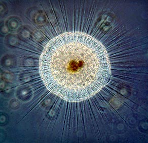
Other adult fresh-water, multicellular organism I first saw is a magnificent rotifer on the muck and it was showing off its long finger cilia. First spooky as it looks but it contains 3 layers of digestion, muscle, and invertebrate shells. It has a full digestive system and it uses the long finger cilia to move food into its mouth and manipulate the water around them in order to move. The fresh-water microorganism is known as Limnias, Setphanoceros Eichornii (Plaskitt 219). See Mp4: Limnias at below. Compressed in 8.43 megabytes. Time: 1 minute, 29 seconds.
MP4: LIMNIAS
There are more rotifers on Plant A & B. There are 4 types of rotifers found in my glass tank. Other rotifer I saw in the tank is known as Philodina (Plaskitt 194). There are trap planet, diatoms - Bacillariophyta (Rainis et cal 116), Euglena helicoideus (Rainis et cal 283), desmids - Conochiloidses & Conochilus (D. Smith 179), Spondylosium (G. Smith 333) in the tank.
See Philodina in the MP4 file (Quickplayer 7) at below and it is compressed in 13 megabytes. It is at a magnification of medium and high power with a playing time of 1 minute, 41 seconds. Philodina: You can see the movement of short cilia on the margins of its mouth when it comes out and spread its mouth (G. Smith 192).
MP4: Philodina
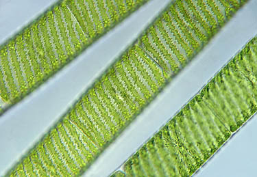
Other good-ole organisms are doing their job to take care of my glass tank: see figure of diatoms and spondylosium at the bottom. Diatoms, to specifically identify it are difficult, but I think it as Mastogloia from the book, The Fresh-Water Algae (Prescott 243). It moves to left and right in slow motion or none at all. It is autotrophic in nature.

Fig. Diatoms & Spondylosium
REFERENCES
McFarland. (2009) General Botany 111 2009. Retrieved data by 27, Oct. 2009. http://botany1112009.blogspot.com/
Plaskitt, F.J.W. (1926). Microscopic Fresh-Water Life. Chapman & Hall, LTD: London. http://www.archive.org/details/microscopicfresh029632mbp. 194, 219
Prescott, G.W. (1964). The Fresh-Water Algae. W.M.C. Brown Company Publishers. 243
Rainis, Kenneth G. & Russell, Bruce J. (1996). Guide to Microlife. Franklin Watts: Danbury, Connecticut. 283
Smith, Douglas Grant. (2001). Pennak’s Freshwater invertebrates of the United States 4th ed. John Wiley & Sons, IAC: New York. 179
Smith, Gilbert M. (1950). The Fresh Water Algae of the United States 2nd ed. McGraw Hill book Company: New York. 333